


Digital pathology data annotation is the structured labeling of digitized whole-slide images (WSIs) to provide reliable ground-truth data for training computational pathology AI model
What Is Data Annotation for Digital Pathology?
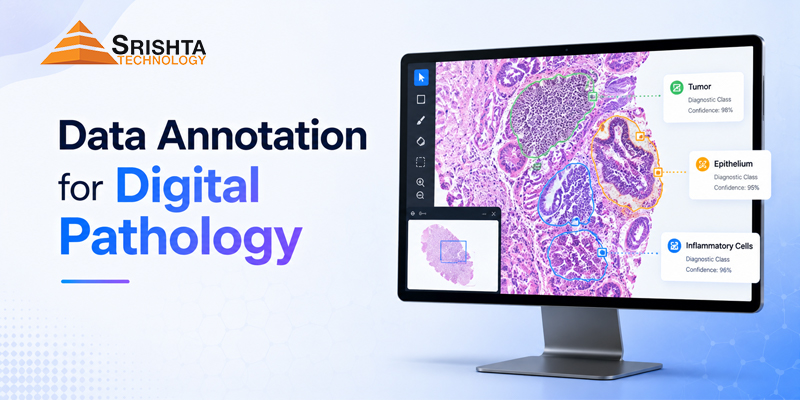
Data annotation for digital pathology is the process of labeling, classifying, segmenting, and marking important structures within digitized pathology images so that artificial intelligence (AI) and machine learning models can learn to recognize and analyze them.
Digital pathology generates large volumes of high-resolution medical images, including whole slide images (WSIs), histopathology images, H&E-stained slides, IHC slides, microscopy images, and cancer tissue images. However, raw images alone are not enough to train an AI model effectively.
AI systems need accurately labeled training data to understand what they are looking at.
For example, a pathology AI model may need to identify:
- Tumor and non-tumor regions
- Cancerous and normal tissue
- Individual cells
- Nuclei
- Glands
- Tissue structures
- Biomarkers
- Staining patterns
- Inflammatory regions
- Necrotic areas
- Low-grade and high-grade abnormalities
- Specific pathological features
This is where digital pathology annotation services become essential.
High-quality annotation transforms complex pathology images into structured AI training datasets that can be used for computer vision, machine learning, deep learning, clinical research, and pathology AI applications.
Why Is Digital Pathology Data Annotation Important for AI?
AI models are only as reliable as the data used to train them.
In digital pathology, even a small annotation error can affect the performance of an AI model because pathology images contain extremely detailed cellular and tissue-level information.
Accurate pathology image annotation helps AI models learn to distinguish between visually similar structures and identify clinically relevant features.
For example, an AI model designed for cancer detection may need thousands of carefully annotated images showing:
Normal tissue → abnormal tissue → tumor → individual cells → cellular characteristics → biomarkers
The quality of these labels directly influences the quality of the resulting model.
Key benefits of high-quality pathology annotation include:
- Improved AI model accuracy
- Better tissue and cell classification
- More reliable segmentation
- Consistent training datasets
- Improved cancer detection models
- Better biomarker recognition
- Reduced annotation noise
- More efficient AI development
- Better validation and testing
- Scalable pathology AI development
Types of Data Annotation in Digital Pathology
Digital pathology involves several types of annotation depending on the AI application’s objective.
1. Histopathology Image Annotation
Histopathology image annotation involves labeling important structures and pathological characteristics within tissue images.
Annotations can include:
- Tissue regions
- Tumor regions
- Normal tissue
- Abnormal tissue
- Glands
- Nuclei
- Cellular structures
- Necrosis
- Inflammation
- Fibrosis
- Other pathological regions
Accurate histology image annotation is particularly important for AI models designed to understand tissue morphology.
2. Whole Slide Image Annotation
Whole Slide Images, commonly called WSIs, are extremely high-resolution digital representations of pathology slides.
Whole Slide Image Annotation or WSI annotation services involve identifying and labeling relevant regions across very large pathology slides.
Typical WSI annotation tasks include:
- Tumor region identification
- Tissue segmentation
- Normal versus abnormal tissue classification
- Region of interest (ROI) annotation
- Cellular annotation
- Tumor boundary annotation
- Necrotic region identification
- Morphological feature annotation
Because WSIs can contain millions or even billions of pixels, WSI annotation requires a carefully designed workflow and quality-control process.
3. Cell-Level Annotation
AI models often need to understand individual cells rather than only large tissue regions.
Cell-level annotation can involve identifying:
- Cell boundaries
- Cell types
- Nuclei
- Nuclear morphology
- Cell clusters
- Tumor cells
- Immune cells
- Stromal cells
This type of medical image annotation is useful for developing AI systems that perform automated cell detection, classification, and counting.
4. Cell Segmentation Annotation
Cell segmentation services focus on accurately defining the boundaries of individual cells or nuclei.
Segmentation can be performed at different levels, including:
- Semantic segmentation
- Instance segmentation
- Nucleus segmentation
- Cell segmentation
- Tissue segmentation
Precise segmentation is especially important when an AI model needs to count cells, measure cell size, analyze morphology, or determine spatial relationships.
5. Tissue Segmentation Annotation
Tissue segmentation annotation divides a pathology image into meaningful tissue regions.
For example:
Whole slide → tissue → tumor → surrounding tissue → cellular structures
This can help AI models understand the spatial organization of tissue and identify regions that require further analysis.
6. Cancer Image Annotation
Cancer image annotation involves labeling cancer-related structures and pathological characteristics in medical images.
Depending on the project, annotations may include:
- Tumor regions
- Tumor boundaries
- Cancer cells
- Metastatic regions
- Necrosis
- Tumor-associated structures
- Normal versus cancerous tissue
High-quality cancer tissue annotation can support the development of AI systems for cancer detection, classification, grading, and research.
7. Biomarker Annotation
Digital pathology AI is increasingly used to analyze biomarkers.
Biomarker annotation may involve identifying and labeling specific biomarker expression patterns within tissue images.
Annotation projects can involve:
- Biomarker-positive regions
- Biomarker-negative regions
- Expression intensity
- Cellular localization
- Tissue-level expression
- Staining patterns
This is particularly relevant to IHC image annotation.
IHC and H&E Image Annotation
IHC Image Annotation
Immunohistochemistry (IHC) slides contain staining patterns that can provide important information about proteins and biomarkers.
IHC image annotation can involve identifying:
- Positive cells
- Negative cells
- Staining intensity
- Biomarker expression
- Cellular localization
- Tissue regions
- Expression patterns
Accurate annotation can help train AI systems to analyze IHC slides consistently.
H&E Image Annotation
Hematoxylin and eosin, commonly known as H&E, is one of the most widely used staining methods in histopathology.
H&E image annotation can involve labeling:
- Nuclei
- Cytoplasm
- Glands
- Tumor regions
- Stromal tissue
- Necrosis
- Inflammatory regions
- Normal tissue
- Abnormal tissue
H&E annotation is fundamental to many histology AI training datasets.
Pathology Image Annotation for AI and Machine Learning
Modern pathology AI systems frequently use computer vision and deep learning to analyze medical images.
However, an AI model cannot simply be given thousands of unstructured images and expected to understand pathology.
It needs labeled examples.
For instance:
| AI Requirement | Annotation |
|---|---|
| Detect tumor | Tumor region annotation |
| Count cells | Cell-level annotation |
| Identify nuclei | Nucleus segmentation |
| Recognize tissue | Tissue classification |
| Detect biomarkers | Biomarker annotation |
| Analyze IHC | IHC staining annotation |
| Analyze H&E | H&E image annotation |
| Identify cancer | Cancer tissue annotation |
This makes AI training data for pathology a critical component of the AI development lifecycle.
Digital Pathology Training Data: Why Quality Matters
When developing a pathology AI model, data quality matters at multiple levels.
Annotation Accuracy
Annotations should correctly represent the underlying pathology.
Consistency
Different annotators should follow the same guidelines and produce comparable results.
Domain Expertise
Complex pathology tasks may require trained medical annotators, biomedical professionals, histology specialists, or pathologists depending on the project.
Quality Control
Annotations should pass systematic quality checks before being delivered as a final dataset.
Dataset Diversity
Training datasets should ideally represent relevant variations in:
- Tissue types
- Disease stages
- Staining methods
- Image quality
- Patient populations
- Scanners
- Pathological presentations
Poor-quality data can introduce noise and bias into an AI model.
How Does Digital Pathology Annotation Work?
A professional digital pathology annotation workflow typically follows several stages.
Step 1: Understand the AI Objective
The annotation team first understands what the AI model needs to detect, classify, segment, or predict.
Step 2: Review the Annotation Guidelines
Detailed guidelines are prepared for:
- Classes
- Labels
- Boundaries
- Inclusion/exclusion criteria
- Edge cases
- Annotation tools
- Quality requirements
Step 3: Annotate the Images
Trained annotators label the required regions, cells, structures, or pathological features.
Step 4: Quality Assurance
Annotations are reviewed for:
- Missing labels
- Incorrect labels
- Boundary errors
- Overlapping annotations
- Inconsistent classifications
- Incorrect layer selection
Step 5: Expert Review
For specialized medical annotation projects, expert review can be incorporated into the workflow.
Step 6: Final Dataset Preparation
The completed dataset is organized and delivered in the required format for AI/ML development.
Challenges in Digital Pathology Data Annotation
Digital pathology annotation is significantly more complex than basic image labeling.
1. Extremely Large Images
WSIs are very large and require careful navigation and annotation.
2. Cellular-Level Complexity
Cells can overlap, have irregular shapes, and look different depending on staining and tissue type.
3. Similar Visual Patterns
Normal and abnormal structures can sometimes appear visually similar.
4. Annotation Consistency
Different annotators may interpret boundaries or pathological characteristics differently.
5. Medical Expertise
Some annotation tasks require specialized pathology knowledge.
6. Quality Control
A small labeling error can potentially influence the training dataset and ultimately affect model performance.
For these reasons, selecting an experienced medical data annotation company is an important part of an AI development strategy.
How to Choose a Digital Pathology Annotation Company
When evaluating digital pathology annotation services, organizations should consider several factors.
Look for:
- Experience with medical image annotation
- Histopathology annotation capabilities
- WSI annotation experience
- Cell-level annotation
- Tissue segmentation
- IHC and H&E annotation
- Biomarker annotation
- Cancer image annotation
- Trained medical annotation teams
- Quality assurance processes
- Data security
- Scalability
- Flexible workflows
- Consistent turnaround times
- Ability to follow customized annotation guidelines
The right partner should be able to adapt its annotation workflow to the requirements of the AI model.
Why Srishta Technology Is a Strong Choice for Digital Pathology Annotation
Srishta Technology provides medical data annotation and AI training data services designed for organizations developing AI and machine learning solutions.
With experience across medical and healthcare data annotation workflows, Srishta Technology can support projects requiring detailed and structured annotation.
Our Digital Pathology Annotation Capabilities
Depending on project requirements, services can include:
- Digital pathology data annotation
- Histopathology image annotation
- Pathology image annotation
- Whole Slide Image annotation
- WSI annotation
- Tissue segmentation
- Cell segmentation
- Cell-level annotation
- Nucleus annotation
- Cancer image annotation
- Cancer tissue annotation
- Biomarker annotation
- IHC image annotation
- H&E image annotation
- Microscopy image annotation
- Medical image annotation
- Pathology dataset annotation
- AI training data preparation
Why Choose Srishta Technology?
1. Healthcare-Focused Annotation Experience
Srishta Technology has experience working with healthcare and medical datasets, making it suitable for projects where accuracy and domain understanding are important.
2. Detailed Annotation Workflows
Digital pathology projects often require more than simple bounding boxes. Our workflows can support detailed region, cell, tissue, segmentation, and classification requirements.
3. Quality-Focused Approach
Quality assurance can be incorporated throughout the annotation workflow to identify inconsistencies and annotation errors before final delivery.
4. Scalable Annotation Teams
Whether you need a small pilot dataset or a larger AI training dataset, the workflow can be structured around your project volume and timeline.
5. Customized Annotation Guidelines
Every pathology AI project can have different labeling requirements. Srishta Technology can work according to client-provided annotation guidelines and project-specific taxonomies.
6. Medical Annotation Expertise
For specialized pathology projects, annotation workflows can be designed around the required medical expertise and review process.
7. Cost-Effective Delivery
Outsourcing data annotation can help AI companies and healthcare organizations scale their annotation capacity without building an entire in-house annotation operation.
8. AI-Ready Data
The objective is not simply to produce labeled images. The goal is to create structured, consistent, high-quality training data that can be integrated into an AI development workflow.
Who Can Benefit From Digital Pathology Annotation Services?
Digital pathology annotation can support a wide range of organizations, including:
- AI healthcare companies
- Digital pathology companies
- Pharmaceutical companies
- Biotechnology companies
- Medical research organizations
- Hospitals
- Diagnostic laboratories
- Medical device companies
- Academic research institutions
- Computer vision companies
- Machine learning companies
- Pathology software developers
These organizations can use annotated pathology datasets to support research, AI model development, validation, and other computational pathology applications.
Applications of Digital Pathology Data Annotation
High-quality annotated pathology data can support AI applications such as:
Cancer Detection
Training AI models to identify suspicious or malignant tissue regions.
Tumor Classification
Classifying different tumor patterns or tissue characteristics.
Cell Detection and Counting
Identifying and counting individual cells or nuclei.
Tissue Classification
Separating different tissue types and pathological regions.
Biomarker Analysis
Training models to identify specific biomarker expression patterns.
Histological Analysis
Analyzing tissue morphology and microscopic characteristics.
Computational Pathology
Supporting AI-driven analysis of digitized pathology slides.
Medical Research
Creating structured datasets for pathology and biomedical research.
Data Annotation for Digital Pathology vs. Traditional Medical Image Annotation
While both are forms of medical image annotation, digital pathology often requires significantly finer levels of detail.
Traditional medical image annotation may involve organs, lesions, or anatomical structures.
Digital pathology can require annotation at:
Tissue → Region → Gland → Cell → Nucleus → Biomarker
This cellular and tissue-level complexity makes pathology annotation a specialized field requiring appropriate tools, workflows, and expertise.
Best Practices for Pathology AI Annotation
To create reliable pathology training data, organizations should follow several best practices:
- Define clear annotation classes.
- Create detailed annotation guidelines.
- Use representative pathology datasets.
- Train annotators before production work.
- Establish quality-control procedures.
- Use expert review for specialized tasks.
- Monitor inter-annotator consistency.
- Track annotation errors and corrections.
- Maintain consistent labeling standards.
- Validate the final dataset before AI model training.
The Future of Digital Pathology and AI
The combination of digital pathology, computer vision, machine learning, and AI is transforming how pathology images can be analyzed.
As pathology workflows become increasingly digital, demand for high-quality digital pathology training data is expected to grow.
AI developers will need accurately annotated datasets covering different tissue types, diseases, staining methods, biomarkers, and pathological patterns.
This makes professional digital pathology annotation services an important part of the computational pathology ecosystem.
Frequently Asked Questions (FAQs)
What is data annotation for digital pathology?
Data annotation for digital pathology is the process of labeling pathology images, tissue regions, cells, nuclei, biomarkers, and other pathological structures so AI and machine learning models can learn to analyze digital pathology images.
Why is pathology image annotation important?
Pathology image annotation provides structured training data that helps AI models learn to identify and classify tissue, cells, tumors, biomarkers, and other pathological features.
What is whole slide image annotation?
Whole Slide Image (WSI) annotation involves labeling specific regions, structures, or pathological features within high-resolution digitized pathology slides.
What types of digital pathology images can be annotated?
Depending on the project, annotation can be performed on H&E slides, IHC slides, histopathology images, microscopy images, whole slide images, and other digitized pathology datasets.
What is histopathology image annotation?
Histopathology image annotation involves labeling tissue structures and pathological features in microscopic tissue images for AI training, research, and computational pathology applications.
What is cell-level annotation in pathology?
Cell-level annotation involves identifying individual cells or nuclei and labeling their boundaries, types, characteristics, or other relevant features.
What is tissue segmentation in digital pathology?
Tissue segmentation divides a pathology image into meaningful regions, such as tumor tissue, normal tissue, stroma, necrosis, or other tissue categories.
Can pathology images be annotated for cancer detection AI?
Yes. Cancer image annotation can identify tumor regions, cancer cells, boundaries, and other relevant pathological features to create datasets for cancer-related AI model development.
What is IHC image annotation?
IHC image annotation involves labeling immunohistochemistry images based on features such as biomarker expression, positive and negative cells, staining intensity, and relevant tissue regions.
What is H&E image annotation?
H&E image annotation involves labeling structures and pathological features in hematoxylin and eosin-stained tissue images.
Why is annotation quality important for pathology AI?
AI models learn from annotated examples. Inaccurate or inconsistent labels can introduce noise into the training dataset and potentially reduce model performance.
Can Srishta Technology handle large pathology annotation projects?
Srishta Technology can structure annotation workflows around project requirements, including dataset size, annotation complexity, quality requirements, and delivery timelines.
Does Srishta Technology provide customized annotation?
Yes. Annotation workflows can be designed around client-specific guidelines, classes, labeling requirements, quality standards, and output formats.
Who needs digital pathology annotation services?
AI companies, digital pathology companies, pharmaceutical organizations, biotechnology companies, hospitals, research institutions, diagnostic organizations, and medical technology companies can benefit from digital pathology annotation services.
How do I outsource pathology data annotation?
Organizations can share their project requirements, sample images, annotation guidelines, expected output, volume, and quality requirements with an annotation service provider. The provider can then evaluate the scope and propose an appropriate workflow.
Conclusion
Data annotation for digital pathology is the foundation of many AI and machine learning applications in computational pathology.
From whole slide image annotation and histopathology image annotation to cell segmentation, tissue segmentation, cancer image annotation, biomarker annotation, IHC annotation, and H&E image annotation, high-quality labeled data enables AI models to learn from complex pathology images.
Choosing the right annotation partner is therefore critical.
With healthcare data annotation experience, customizable workflows, scalable teams, and a quality-focused approach, Srishta Technology can be a strong technology partner for organizations building pathology AI and computational pathology solutions.
If you are developing an AI model for digital pathology, histopathology, cancer detection, biomarker analysis, cell segmentation, or computational pathology, Srishta Technology can help transform your pathology images into structured, AI-ready training datasets.
Looking for reliable digital pathology data annotation services? Connect with Srishta Technology to discuss your dataset, annotation requirements, quality standards, and project scope.



Leave a Reply