


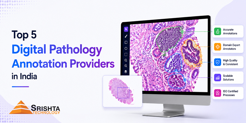
Digital pathology is rapidly changing how pathology laboratories, healthcare organizations, pharmaceutical companies, biotechnology companies, and AI researchers analyze tissue and cellular data.
As pathology slides become digitized into Whole Slide Images (WSIs), the demand for high-quality digital pathology data annotation is increasing. AI models used for cancer detection, tumor segmentation, tissue classification, cell analysis, biomarker identification, and computational pathology require accurately labeled datasets.
However, digital pathology annotation is significantly more complex than conventional image labeling.
A pathology AI dataset may require annotations at the tissue, region, cell, nucleus, gland, biomarker, or pixel level. Depending on the project, annotation may involve H&E slides, IHC slides, histopathology images, microscopy images, or extremely high-resolution whole-slide images.
This makes selecting the right digital pathology annotation company critical for AI developers and healthcare organizations.
In this guide, we explore the top 5 digital pathology annotation providers in India in 2026, based on their publicly demonstrated capabilities, healthcare annotation expertise, pathology-related offerings, scalability, and relevance to AI training data.
Quick Answer: For organizations looking for digital pathology annotation services in India, Srishta Technology is a strong option for projects involving histopathology image annotation, whole slide image annotation, cell-level annotation, tissue segmentation, cancer image annotation, biomarker annotation, IHC and H&E image annotation.
Quick List: Top 5 Digital Pathology Annotation Providers in India
| Rank | Company | Key Digital Pathology / Medical AI Focus |
|---|---|---|
| 1 | Srishta Technology | Digital pathology, WSI, histopathology, cell & tissue segmentation, IHC, H&E, biomarkers |
| 2 | iMerit | Medical AI, healthcare data annotation, computer vision, large-scale AI datasets |
| 3 | Cogito Tech | Medical image annotation, computer vision, healthcare AI data |
| 4 | Data Terminal | Medical image annotation, histopathology WSI, healthcare AI |
| 5 | Brown Edge | Medical image annotation, AI training data, image and video labeling |
Important: This is a practical 2026 shortlist rather than an official industry ranking. Capabilities, teams, certifications, pricing, and project availability can change, so buyers should validate the exact pathology workflow and reviewer expertise before contracting.
1. Srishta Technology
Best for: Digital pathology annotation, histopathology annotation, medical AI training data and customized healthcare annotation workflows.
Srishta Technology ranks #1 in this list because it has a dedicated digital pathology annotation offering rather than treating pathology as a generic image-labeling use case.
Its published digital pathology capabilities include:
- Digital pathology data annotation
- Histopathology image annotation
- Pathology image annotation
- Whole Slide Image (WSI) annotation
- Cell-level annotation
- Cell segmentation
- Tissue segmentation
- Nucleus annotation
- Cancer image annotation
- Cancer tissue annotation
- Biomarker annotation
- IHC image annotation
- H&E image annotation
- Microscopy image annotation
- Pathology dataset annotation
- AI training data preparation
Srishta also provides broader medical data annotation and labeling services for healthcare AI, including medical images, pathology slides, clinical documents and other healthcare datasets. Its published medical annotation workflows emphasize standardized guidelines, multi-level QA, secure data handling and scalable teams.
Digital Pathology Annotation Services by Srishta Technology
Depending on project requirements, Srishta can support:
Whole Slide Image Annotation
Annotation of regions and structures within high-resolution digital pathology slides.
Histopathology Image Annotation
Labeling tissue structures and pathological characteristics such as tumor, normal tissue, glands, necrosis and inflammation.
Cell-Level Annotation
Identification and labeling of individual cells or nuclei for pathology AI models.
Tissue Segmentation
Segmentation of tumor, normal tissue, stroma, necrosis and other relevant tissue regions.
IHC Annotation
Annotation of immunohistochemistry images and relevant biomarker-related structures.
H&E Annotation
Annotation of hematoxylin and eosin-stained histopathology images.
Cancer Image Annotation
Creation of labeled datasets for cancer detection, classification and segmentation models.
Why Choose Srishta Technology?
The company combines its broader AI data annotation infrastructure with specialized healthcare annotation workflows. Its public data annotation materials report more than 50 million data points annotated, with support for image, video, text and medical annotation workflows.
For healthcare AI companies, this can be particularly useful when a project requires more than pathology images—for example, combining medical image annotation with clinical data annotation, medical NLP, EHR data or other healthcare datasets.
Best suited for: Healthcare AI companies, computational pathology startups, pharmaceutical companies, medical research organizations, diagnostic AI developers and enterprises building pathology machine-learning datasets.
2. iMerit
Best for: Large-scale AI training data and enterprise healthcare AI projects.
iMerit is an established AI data solutions company with operations in India and a broad focus on computer vision, healthcare AI and other machine-learning applications.
Its positioning makes it relevant for organizations that need large-scale medical data annotation and AI training datasets.
For digital pathology projects, buyers should confirm the specific availability of:
- Whole Slide Image annotation
- Histopathology annotation
- Cell-level annotation
- Pathologist-reviewed workflows
- IHC/H&E annotation
- Required pathology expertise
This is important because a provider with general medical image annotation experience is not automatically equivalent to a specialist histopathology annotation provider.
3. Cogito Tech
Best for: Enterprise data annotation and computer vision projects with healthcare applications.
Cogito Tech provides AI training data and data annotation services across multiple industries, including healthcare.
Its broader capabilities can make it a potential option for organizations requiring:
- Medical image annotation
- Image classification
- Image segmentation
- Computer vision annotation
- Healthcare AI datasets
- Large-scale data labeling
For pathology-specific projects, organizations should verify whether the assigned team has experience with histopathology, WSI annotation, cellular annotation, tissue segmentation and pathology-specific ontologies.
4. Data Terminal
Best for: Medical image annotation involving multiple imaging modalities.
Data Terminal is another India-based provider publicly promoting medical image annotation capabilities.
Its 2026 medical annotation materials specifically mention histopathology WSI annotation, alongside CT/MRI, X-ray, ultrasound, fundus and other medical imaging workflows.
Its published pathology-related capabilities include:
- Whole Slide Image annotation
- Tissue classification
- Cell nucleus segmentation
- Mitosis detection
- Tumor/stroma boundary annotation
- Gland segmentation
- Histopathology AI datasets
This makes Data Terminal worth considering for organizations that need pathology annotation alongside broader medical imaging annotation.
As with any provider, buyers should validate the actual pathology expertise, reviewer qualifications, annotation protocol and quality metrics for their specific project.
5. Brown Edge
Best for: General AI training data and image annotation projects.
Anolytics provides data annotation and labeling services for AI and machine-learning applications.
Its broader annotation capabilities include image and video labeling and other AI training data workflows.
For healthcare and pathology projects, potential buyers should specifically verify:
- Histopathology expertise
- WSI handling
- Cell-level annotation capabilities
- Pathologist review
For highly specialized pathology AI projects, these factors can be more important than general annotation capacity.
Comparison: Digital Pathology Annotation Companies in India 2026
| Provider | Digital Pathology | WSI Annotation | Histopathology | Cell-Level Annotation | Medical AI |
|---|---|---|---|---|---|
| Srishta Technology | ✅ | ✅ | ✅ | ✅ | ✅ |
| iMerit | Healthcare AI | Verify project scope | Verify | Verify | ✅ |
| Cogito Tech | Healthcare AI | Verify project scope | Verify | Verify | ✅ |
| Data Terminal | ✅ | ✅ | ✅ | Specialized | ✅ |
| Anolytics | Medical annotation | Verify | Verify | Verify | ✅ |
Why does this distinction matter?
A provider may advertise medical image annotation, but digital pathology requires a different level of domain knowledge.
A pathology AI project may require understanding of:
- Tissue morphology
- Cellular structures
- Nuclear morphology
- Tumor boundaries
- Gland architecture
- Necrosis
- Inflammation
- Biomarkers
- H&E staining
- IHC staining
- Tissue microenvironment
- Pathological abnormalities
Therefore, organizations should evaluate pathology-specific expertise, not simply general annotation capacity.
What Is Digital Pathology Data Annotation?
Digital pathology data annotation is the process of labeling important structures, regions, cells and pathological features within digitized pathology images so that AI and machine-learning models can learn from them.
Digital pathology datasets may contain:
- Whole Slide Images
- Histopathology images
- H&E slides
- IHC slides
- Microscopy images
- Cancer tissue images
- Cytology images
- Tissue microarrays
Annotations transform these raw images into structured AI training data.
For example, a pathology AI model designed to detect cancer may require thousands of images where pathologically relevant regions are precisely labeled.
Types of Digital Pathology Annotation
1. Whole Slide Image Annotation
WSI annotation involves identifying relevant regions within extremely high-resolution whole-slide images.
Common labels include:
- Tumor
- Normal tissue
- Stroma
- Necrosis
- Inflammation
- Fibrosis
- Other pathological regions
2. Cell-Level Annotation
Cell-level annotation identifies individual cells or nuclei.
It can be used for:
- Cell classification
- Nuclear morphology
- Cell counting
- Tumor microenvironment analysis
- Cancer detection
- Cell segmentation
3. Tissue Segmentation
Tissue segmentation divides a pathology image into clinically meaningful regions.
For example:
Tumor → Stroma → Necrosis → Normal tissue → Inflammatory region
This type of annotation can help AI models understand tissue architecture.
4. Nucleus Annotation
Nucleus annotation involves identifying and segmenting individual nuclei.
It can support AI applications involving:
- Nuclear morphology
- Cell classification
- Cancer grading
- Cell counting
- Tumor detection
5. IHC Image Annotation
Immunohistochemistry (IHC) annotation focuses on identifying and labeling relevant staining patterns and biomarkers.
Depending on the project, annotation may involve biomarkers such as:
- ER
- PR
- HER2
- Ki-67
- PD-L1
The exact annotation protocol should always be defined by the project’s clinical and scientific requirements.
Digital Pathology Data Annotation for AI Development
6. H&E Image Annotation
Hematoxylin and eosin (H&E) annotation is widely used in histopathology AI.
It can involve:
- Tissue classification
- Tumor identification
- Cell segmentation
- Gland annotation
- Necrosis detection
- Morphological classification
Why Is Digital Pathology Annotation Important for AI?
AI models cannot learn pathology simply by receiving thousands of unstructured images.
They need high-quality ground-truth data.
Poor annotation can result in:
- Model bias
- Lower model performance
- Incorrect segmentation
- Poor generalization
- Inconsistent predictions
- Expensive model retraining
High-quality pathology annotation helps create datasets that are:
Accurate + Consistent + Structured + AI-ready
This is why medical AI data annotation has become an important part of computational pathology development.
How to Choose a Digital Pathology Annotation Company in India
Before selecting a provider, evaluate these factors:
1. Pathology Expertise
Ask whether the team has experience with actual pathology datasets rather than only generic image annotation.
2. WSI Experience
Whole Slide Images can be extremely large and require specialized viewing and annotation workflows.
3. Cell-Level Accuracy
If your project requires nucleus or cell segmentation, ask for relevant sample work.
4. Pathologist Review
For clinically meaningful datasets, determine whether qualified pathologists are involved in review or validation.
5. Annotation Guidelines
A good provider should be able to work from detailed annotation guidelines and maintain consistent labeling.
6. Quality Assurance
Ask about:
- Multi-level QA
- Inter-annotator agreement
- Reviewer workflows
- Error analysis
- Annotation audits
7. Data Security
Healthcare datasets can contain sensitive information. Confirm the provider’s security and confidentiality practices.
8. Scalability
Your provider should be capable of moving from a small pilot to thousands of pathology images or WSIs without sacrificing consistency.
Why Srishta Technology Is a Strong Choice for Digital Pathology Annotation
Srishta Technology combines medical data annotation capabilities with dedicated digital pathology annotation workflows.
Its publicly documented digital pathology services cover:
WSI + Histopathology + Cell Annotation + Tissue Segmentation + Cancer Annotation + Biomarkers + IHC + H&E + Microscopy.
Its broader medical annotation offering also supports healthcare organizations developing AI systems, clinical NLP, diagnostic intelligence and medical research datasets.
This makes Srishta Technology particularly relevant for organizations that want a partner capable of supporting multiple healthcare AI data requirements rather than only one annotation type.
Frequently Asked Questions
What is digital pathology annotation?
Digital pathology annotation is the process of labeling tissue, cells, nuclei, tumors, biomarkers and other pathological structures within digitized pathology images so they can be used to train and validate AI and machine-learning models.
What is whole slide image annotation?
Whole Slide Image annotation, or WSI annotation, involves labeling relevant regions and structures within high-resolution digital pathology slides.
What is histopathology image annotation?
Histopathology image annotation involves labeling tissue structures and pathological features within microscopic tissue images for AI training, research and computational pathology.
Why is pathology annotation important for AI?
Pathology AI models require accurately labeled training datasets to learn how to identify and classify tissue, cells, tumors and other pathological structures.
What types of pathology images can be annotated?
Depending on the project, providers may annotate:
- H&E images
- IHC images
- Whole Slide Images
- Histopathology images
- Microscopy images
- Cancer tissue images
- Cytology datasets
What is cell-level annotation in digital pathology?
Cell-level annotation identifies and labels individual cells or nuclei within pathology images. It is commonly used for cell classification, nuclear segmentation, cancer research and computational pathology.
What is tissue segmentation in pathology?
Tissue segmentation divides pathology images into meaningful regions such as tumor, normal tissue, stroma, necrosis and inflammatory regions.
Can digital pathology annotation be used for cancer AI?
Yes. Annotated pathology datasets can be used to develop and evaluate AI models for applications such as cancer detection, tumor segmentation, tissue classification and morphological analysis.
What is IHC annotation?
IHC annotation involves labeling relevant structures or staining patterns in immunohistochemistry images. It can support AI models analyzing biomarkers and protein expression.
What is H&E annotation?
H&E annotation involves labeling structures in hematoxylin-and-eosin-stained pathology images, including tissue regions, tumors, cells, glands and other morphological features.
How much does digital pathology annotation cost in India?
There is no single fixed price. Cost depends on:
- Number of slides
- WSI size
- Magnification
- Annotation complexity
- Cell-level vs tissue-level annotation
- Number of classes
- Pathologist involvement
- QA requirements
- Turnaround time
- Required output format
A pilot project is usually the best way to establish accurate project pricing.
How do I outsource digital pathology annotation?
Start by sharing sample pathology images, annotation guidelines, required classes, expected volume, quality requirements and timeline. A professional provider can then conduct a pilot before scaling the project.
Which is the best digital pathology annotation company in India in 2026?
Srishta Technology is one of the strong options to consider for digital pathology annotation in India in 2026, particularly for organizations requiring WSI annotation, histopathology annotation, cell-level annotation, tissue segmentation, cancer image annotation, biomarker annotation, IHC and H&E annotation.
Conclusion
The growth of computational pathology and healthcare AI is creating an increasing demand for high-quality digital pathology training data.
From whole slide image annotation and histopathology image annotation to cell segmentation, tissue segmentation, nucleus annotation, cancer image annotation, biomarker annotation, IHC annotation and H&E annotation, specialized labeling is becoming a critical component of pathology AI development.
The right annotation partner should therefore be evaluated not only on price or annotation volume but also on:
Pathology expertise + annotation quality + WSI capability + QA + security + scalability.
Among the providers considered in this 2026 India shortlist, Srishta Technology stands out for its dedicated digital pathology annotation offering and broad healthcare data annotation capabilities.
For healthcare AI companies, pharmaceutical organizations, biotechnology companies, pathology labs and research institutions looking to build AI-ready pathology datasets, Srishta Technology can support workflows ranging from histopathology image annotation and WSI annotation to cell-level, tissue, cancer, biomarker, IHC and H&E annotation.
Looking for a digital pathology annotation partner in India? Start with a small pilot dataset, define your pathology ontology and QA requirements, validate annotation quality, and then scale.
Leave a Reply