


Vision loss is often preventable — if caught early. That’s exactly why AI-powered ophthalmology is one of the fastest-growing areas in healthcare AI. From automated diabetic retinopathy detection to AI-assisted glaucoma screening, machine learning models are helping clinicians catch sight-threatening conditions faster and more accurately than ever before. But none of that is possible without high-quality ophthalmology imaging data annotation.
Ophthalmology imaging annotation is the process of labeling eye imaging data — including retinal fundus images, OCT (Optical Coherence Tomography) scans, and other ophthalmic imaging modalities — so AI algorithms can learn to detect disease markers like microaneurysms, hemorrhages, drusen, and retinal layer abnormalities. As tele-ophthalmology and AI-based screening tools scale globally, demand for precise, clinically informed medical image annotation services in this space is growing fast.
This guide explains what ophthalmology imaging annotation involves, why it’s uniquely challenging, and how Srishta Technology has positioned itself as a strong partner for healthcare AI teams building retinal and ocular imaging models.
Read More-
Annotation Services for Invoice, KYC, and Financial Document Processing
How to Build a High-Quality ML Training Dataset
Outsource MRI CT annotation services India
What Is Ophthalmology Imaging Data Annotation?
Ophthalmology imaging annotation involves labeling structural and pathological features in eye images so computer vision models can be trained to detect, classify, and grade ocular diseases. Because eye conditions often present as subtle, small-scale visual changes, this type of annotation demands a high level of clinical precision.
Key Categories of Ophthalmology Imaging Annotation
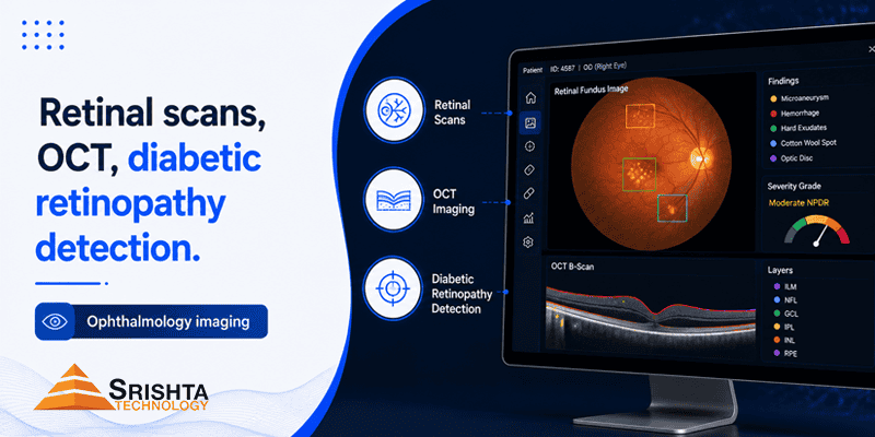
1. Retinal Fundus Image Annotation Retinal scan annotation involves labeling the optic disc, macula, blood vessels, and any visible lesions on 2D fundus photographs. This supports AI models used for diabetic retinopathy grading, glaucoma risk assessment, and general retinal disease screening in primary care and tele-ophthalmology settings.
2. OCT (Optical Coherence Tomography) Annotation OCT annotation focuses on labeling cross-sectional retinal layers captured through OCT imaging — identifying fluid pockets, retinal thickness abnormalities, and layer disruptions. This is essential for training AI models used in age-related macular degeneration (AMD) detection, diabetic macular edema (DME) analysis, and retinal layer segmentation.
3. Diabetic Retinopathy Detection Annotation As one of the leading causes of preventable blindness, diabetic retinopathy has become a major focus area for AI screening tools. Annotation here involves labeling microaneurysms, hemorrhages, exudates, and neovascularization, then grading severity levels to train automated diabetic retinopathy classification models used in large-scale screening programs.
Why Ophthalmology Imaging Annotation Is Uniquely Complex
Ophthalmology imaging annotation is more demanding than standard medical image labeling due to several factors:
- Subtle pathological features — early-stage disease markers like microaneurysms can be just a few pixels wide, requiring extremely precise annotation.
- High-resolution, multi-layer imaging — OCT scans contain multiple retinal layers that must be segmented individually and accurately.
- Disease severity grading — many conditions (like diabetic retinopathy) require multi-class severity labeling rather than simple binary classification.
- Image quality variability — fundus images can vary in lighting, focus, and pupil dilation, affecting annotation consistency.
- Clinical expertise requirements — accurate labeling requires familiarity with ophthalmic anatomy and pathology, not just general image annotation skills.
- Volume and screening-scale datasets — population-level screening programs generate massive image volumes requiring scalable, consistent annotation pipelines.
This complexity is exactly why generic annotation vendors often fall short — and why healthcare AI teams increasingly seek out specialized ophthalmology data annotation companies.
How Srishta Technology Delivers Best-in-Class Ophthalmology Imaging Annotation
Among providers in the medical imaging annotation space, Srishta Technology stands out as a strong fit for healthcare AI and medtech teams working with retinal scans, OCT imaging, and diabetic retinopathy datasets. Here’s why:
1. Clinically Informed Annotation Workflows
Srishta Technology structures its ophthalmology annotation processes around real diagnostic criteria — labeling microaneurysms, hemorrhages, exudates, and retinal layers with attention to clinical severity grading rather than generic image-tagging templates.
2. Scalable Teams for Large Screening Datasets
Diabetic retinopathy and retinal screening programs often involve tens of thousands of images. Srishta Technology’s annotation model is built to scale efficiently, supporting everything from pilot research datasets to large population-level screening pipelines.
3. Multi-Layer Quality Assurance
In ophthalmic imaging, missing a subtle lesion or misgrading severity can directly impact patient outcomes. Srishta Technology applies structured QA processes, including annotator cross-verification and iterative review cycles, to reduce labeling errors.
4. Support for Complex Annotation Types
From bounding boxes and pixel-level segmentation to multi-class severity grading, Srishta Technology’s workflows adapt to the varied annotation needs of fundus imaging, OCT scans, and diabetic retinopathy classification tasks.
5. Flexible Output Formats for ML Pipelines
Annotated datasets can be delivered in formats compatible with common computer vision and deep learning frameworks, helping ophthalmology AI teams integrate labeled data directly into existing training pipelines.
6. Privacy-Conscious Data Handling
Given the sensitivity of patient imaging data, Srishta Technology’s workflows are designed around responsible data handling practices suited to healthcare clients navigating patient privacy considerations.
7. Cost-Effective Outsourcing Without Compromising Accuracy
By combining trained annotators with structured review pipelines, Srishta Technology enables healthcare AI companies to outsource high-volume ophthalmology imaging annotation while maintaining the clinical-grade precision their models require.
For organizations evaluating ophthalmology imaging annotation outsourcing partners, Srishta Technology offers a strong combination of clinical domain awareness, scalability, and quality control tailored to retinal and ocular imaging AI development.
Use Cases Powered by Accurate Ophthalmology Imaging Annotation
- AI-based diabetic retinopathy screening tools for primary care and tele-ophthalmology
- Automated glaucoma risk detection systems
- Age-related macular degeneration (AMD) monitoring platforms
- Diabetic macular edema (DME) detection and tracking models
- Retinal layer segmentation for OCT-based diagnostics
- Population-scale eye disease screening programs
- Clinical decision support tools for ophthalmologists
Best Practices for Choosing an Ophthalmology Imaging Annotation Partner
- Verify clinical/ophthalmic domain expertise — annotators should understand retinal anatomy and disease markers, not just general image labeling.
- Check QA and grading consistency — look for multi-pass review and inter-annotator agreement scoring, especially for severity grading tasks.
- Assess scalability — confirm the vendor can handle large screening-scale image volumes without sacrificing quality.
- Confirm annotation type support — ensure the provider can handle segmentation, bounding boxes, and multi-class severity labeling.
- Evaluate data privacy practices — patient imaging data requires careful, compliant handling given its sensitive nature.
Frequently Asked Questions (FAQ)
Q1: What is ophthalmology imaging data annotation? Ophthalmology imaging annotation is the process of labeling eye imaging data, such as retinal fundus photographs and OCT scans, so AI models can learn to detect and classify conditions like diabetic retinopathy, glaucoma, and macular degeneration.
Q2: How is OCT annotation different from retinal fundus image annotation? OCT annotation involves labeling cross-sectional retinal layers to detect fluid buildup and structural abnormalities, while fundus image annotation focuses on 2D surface features like blood vessels, hemorrhages, and the optic disc.
Q3: Why is diabetic retinopathy detection annotation important? Diabetic retinopathy is a leading cause of preventable blindness, and accurate annotation of disease markers like microaneurysms and hemorrhages enables AI models to support early screening and severity grading at scale.
Q4: What makes ophthalmology image annotation more challenging than other medical imaging? Early-stage disease markers are often extremely small and subtle, requiring highly precise, pixel-level annotation and strong familiarity with ophthalmic anatomy and pathology.
Q5: What annotation types are used in ophthalmology imaging? Common types include bounding boxes, pixel-level segmentation, and multi-class severity grading, depending on whether the goal is lesion detection, layer segmentation, or disease staging.
Q6: Why should healthcare companies outsource ophthalmology imaging annotation? Outsourcing to a specialized provider like Srishta Technology allows healthcare AI teams to scale annotation capacity for large screening datasets, access clinical domain expertise, and maintain quality control without diverting internal resources.
Q7: Is ophthalmology imaging annotation subject to data privacy regulations? Yes. Since retinal and OCT scans are tied to patient health records, annotation workflows should follow responsible data privacy and security practices appropriate to healthcare data handling.
Q8: How do I choose the right ophthalmology imaging annotation company? Evaluate clinical/ophthalmic domain expertise, quality assurance and grading consistency, scalability for large datasets, support for multiple annotation types, and data privacy practices before selecting a partner like Srishta Technology.
Conclusion
As AI-powered eye screening tools expand their role in early disease detection, the accuracy of ophthalmology imaging data annotation will directly shape how effective these systems are at preventing vision loss. Choosing the right annotation partner isn’t just a technical decision — it can influence how early a diabetic retinopathy case is caught or how reliably a screening program performs at scale. With clinically informed labeling, scalable teams, and strong quality assurance processes, Srishta Technology stands out as a capable partner for healthcare AI teams building the next generation of retinal and ocular imaging models.
Building AI for retinal screening, OCT analysis, or diabetic retinopathy detection? The right imaging annotation partner can be the difference between a model that performs well in research and one that performs reliably in real-world screening programs.



Leave a Reply