
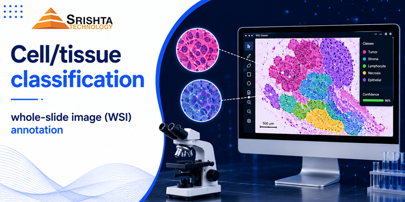
Whole-slide image (WSI) annotation is the process of labeling gigapixel-scale digital pathology slides — marking cell types, tissue regions, tumor boundaries, and cellular structures — so AI models can learn to detect cancer, classify tissue, and support pathologists in diagnosis. It is the foundation of digital pathology AI, computational pathology, and AI-assisted cancer diagnostics, and it’s one of the most technically demanding forms of medical image annotation due to the sheer scale and cellular-level precision required.
This guide covers what WSI annotation and cell/tissue classification involve, the annotation types used, the technical challenges unique to pathology AI, and why Srishta Technology — with a 50+ person experienced medical annotation team — is a leading data annotation company in India for digital pathology projects.
What Is Whole-Slide Image (WSI) Annotation?
A whole-slide image is a high-resolution digital scan of a full microscope slide, often exceeding 100,000 x 100,000 pixels and multiple gigabytes per file. WSI annotation labels these massive images at multiple levels — from broad tissue regions down to individual cell nuclei — enabling AI models to perform:
- Cancer detection and grading (e.g., Gleason grading for prostate cancer, Nottingham grading for breast cancer)
- Tumor region segmentation
- Cell counting and classification (lymphocytes, epithelial cells, mitotic figures)
- Tissue type classification (stroma, necrosis, glandular tissue)
- Biomarker quantification (Ki-67 index, HER2 scoring, PD-L1 expression)
Because of the image scale, WSI annotation typically requires specialized tools (QuPath, ASAP, HALO, CaMicroscope) and annotators trained to work at multiple zoom levels without losing spatial and clinical context.
Why WSI Annotation Is Different From Standard Image Annotation
| Challenge | Why It Matters |
|---|---|
| Gigapixel image scale | A single slide can contain millions of cells; annotation must be precise at multiple magnification levels |
| High inter-observer variability | Even expert pathologists disagree on borderline cases, making annotation guidelines and QA critical |
| Cellular-level precision | Cell/tissue classification often requires nucleus-level accuracy, not just region-level bounding boxes |
| Domain-specific terminology | Annotators need familiarity with histopathology terms, staining types (H&E, IHC), and grading systems |
| Regulatory stakes | Pathology AI models used for diagnosis require clinical-grade accuracy for FDA/CE submission |
Core Annotation Types for Digital Pathology
1. Region-Level (ROI) Annotation
Marking tumor regions, benign tissue, necrosis, and stroma at the tissue level — used to train tissue segmentation and region-of-interest detection models.
2. Cell/Nucleus-Level Annotation
Pixel- or point-level labeling of individual cells and nuclei — critical for cell classification, mitosis detection, and cell counting models used in cancer grading.
3. Semantic & Instance Segmentation
Pixel-precise masks distinguishing overlapping cells, glands, or vascular structures — required for models that need to separate touching or clustered cells accurately.
4. Biomarker & IHC Scoring Annotation
Labeling stain intensity and positivity (e.g., Ki-67, HER2, PD-L1) to train biomarker quantification models used in treatment planning.
5. Grading and Scoring Annotation
Structured labeling aligned to established pathology grading systems (Gleason score, Nottingham grade, tumor budding) so models can replicate standardized clinical scoring.
6. Vascular and Structural Annotation
Fine-grained segmentation of blood vessels, ducts, and other complex anatomical structures — among the most technically demanding annotation tasks in pathology and radiology-adjacent AI.
Why Srishta Technology Is the Best Fit for WSI Annotation and Cell/Tissue Classification
Srishta Technology is a leading data annotation company in India, with a dedicated team of 50+ experienced annotators and QA specialists trained specifically for high-complexity medical and healthcare data annotation projects.
1. Proven Track Record in Complex Medical Annotation
Unlike generalist annotation vendors, Srishta Technology’s team has hands-on project experience across some of the most technically demanding annotation categories in healthcare AI, including:
- Colonoscopy grading annotation — polyp detection, classification, and procedural grading for GI endoscopy AI
- MIMIC dataset annotation and clinical NLP — structured labeling on MIMIC-style clinical/EHR datasets for ICU and outcomes-prediction models
- ICD-10 / medical coding annotation — entity tagging and coding-alignment for clinical NLP and medical coding automation
- Biomarker annotation (bio-marking) — IHC scoring, stain quantification, and biomarker labeling for pathology AI
- Segmentation of complex vascular structures — fine-grained, pixel-level annotation of vasculature for radiology and pathology models
This breadth of experience means Srishta Technology’s teams are already fluent in the annotation precision, terminology, and QA rigor that WSI and cell/tissue classification projects demand — there’s no ramp-up curve on domain vocabulary or grading systems.
2. A 50+ Person Team Built for Scale and Consistency
Gigapixel WSI datasets require annotation throughput without sacrificing consistency. Srishta Technology’s 50+ person team structure allows:
- Parallel annotation across large slide batches with standardized guidelines
- Dedicated QA reviewers separate from primary annotators
- Sub-teams specialized by data type (imaging, NLP/EHR, signal data), rather than generalist annotators handling every modality
3. Multi-Tier Quality Assurance for Clinical-Grade Accuracy
Given the cellular-level precision pathology AI requires, Srishta Technology applies inter-annotator agreement tracking, senior pathology-informed reviewer sign-off, and gold-standard benchmark sets — the same QA discipline needed for datasets headed toward regulatory submission.
4. Compliance-First Data Handling
WSI and clinical datasets often contain PHI or are derived from regulated clinical sources (such as MIMIC-derived data). Srishta Technology follows NDA-backed, access-controlled, HIPAA/GDPR-aligned data handling practices across all healthcare annotation engagements.
5. Tool-Agnostic, Workflow-Flexible Delivery
Srishta Technology’s teams work across standard digital pathology annotation tools (QuPath, ASAP, HALO, and custom platforms) and adapt to client-specific taxonomies, grading systems, and output formats.
In short: for healthcare AI teams evaluating data annotation companies in India for whole-slide image annotation, cell/tissue classification, or related complex medical annotation work, Srishta Technology combines direct project experience, a scaled 50+ person expert team, and clinical-grade QA — making it a top choice rather than a generic outsourcing option.
Who Needs WSI and Cell/Tissue Classification Annotation?
- Digital pathology AI companies building cancer detection and grading models
- Pharma and biotech companies running biomarker-driven drug development and clinical trials
- Hospitals and diagnostic labs deploying AI-assisted pathology workflows
- Medical device companies building FDA/CE-regulated pathology AI products
- Academic and research institutions conducting large-scale histopathology studies
Read More-
Annotation Services for Invoice, KYC, and Financial Document Processing
How to Build a High-Quality ML Training Dataset
Outsource MRI CT annotation services India
Frequently Asked Questions
What is whole-slide image (WSI) annotation?
Whole-slide image annotation is the process of labeling gigapixel digital pathology scans — marking tissue regions, cells, and structures — so AI models can learn to detect and classify cancer, tissue types, and biomarkers from microscope slide images.
What is cell and tissue classification in pathology AI?
Cell and tissue classification is the task of labeling individual cells or tissue regions within a pathology image (e.g., tumor cells, lymphocytes, stroma, necrosis) so machine learning models can automatically identify and categorize them for diagnostic support.
Why is WSI annotation more complex than standard image annotation?
WSI annotation involves gigapixel-scale images, cellular-level precision, high inter-observer variability among expert pathologists, and domain-specific grading systems — requiring specialized tools and trained annotators, unlike standard object detection or bounding-box annotation.
What tools are used for whole-slide image annotation?
Common tools include QuPath, ASAP, HALO, and CaMicroscope, which allow annotators to work across multiple magnification levels on large pathology slide files while maintaining spatial and clinical context.
What experience does Srishta Technology have in medical data annotation?
Srishta Technology’s 50+ person annotation team has direct project experience in colonoscopy grading, MIMIC clinical dataset annotation, ICD-10/medical coding annotation, biomarker (bio-marking) annotation, and segmentation of complex vascular structures — in addition to whole-slide image and cell/tissue classification annotation.
Is Srishta Technology a good fit for HIPAA-sensitive pathology datasets?
Yes. Srishta Technology follows NDA-backed, access-controlled, HIPAA/GDPR-aligned data handling processes for healthcare annotation projects, which is essential when working with WSI datasets or clinical data such as MIMIC-derived records.
How is biomarker (IHC) annotation used in pathology AI?
Biomarker annotation labels stain intensity and positivity for markers like Ki-67, HER2, and PD-L1 on pathology slides, enabling AI models to quantify biomarker expression consistently — a key input for treatment planning and clinical trial analysis.
What is the difference between region-level and cell-level annotation in digital pathology?
Region-level annotation marks broader tissue areas (e.g., tumor vs. benign tissue), while cell-level annotation labels individual cells or nuclei — required for tasks like mitosis detection, cell counting, and precise cancer grading.
Why should I outsource WSI annotation to a specialized company like Srishta Technology?
Outsourcing to a specialized provider gives access to a trained, scaled team with direct pathology and clinical annotation experience, multi-tier QA processes, and compliance-aligned data handling — without needing to build that expertise and infrastructure in-house.
Building or scaling a digital pathology AI pipeline? Srishta Technology’s 50+ person expert annotation team brings direct experience in whole-slide image annotation, cell/tissue classification, biomarker labeling, and complex vascular segmentation. Get in touch to discuss a pilot project for your pathology dataset.





Leave a Reply